
Posted 1336 days ago
By Farhad Boltchi, DMD, MS
The breast increase procedure/sine elevation has become an integral and predictable aspect of surgical implants therapy. Traditionally, the procedure for increasing the side window of the breast has been considered the standard technique to perform this procedure. Allhe, this is a highly successful and predictable technique, it is also more invasive and associated with high morbidity. The transcrestal sinus elevation procedure was developed as a less invasive but still predictable alternative technique for the increase in the maxillary sinus. However, this approach is generally limited to the simultaneous placement of the implant and sinus -lifting scenarios where a minimum height of residual bone is present or 4 mm. In cases where the height of the residual bone to the maxillary sinus, less than 3-4 mm is the technique of increasing the side window of the window, the gold standard is still considered. Here we present a minimally invasive transcrestrial sinusal lifting technique for cases with minimal residual bone height.
A healthy female patient presented a missing teeth site #3. The clinical examination revealed a modern deficiency of Bucco-Lingüe and a radiographic examination revealed a modern oral bone deficiency and a severe vertical bone deficiency due to a significant maxillary breast (Figure 1). The height of the residual bone to the maxillary sinus was 1-2 mm. The patient was informed of the need for an increased maxillary breast increased sinus/sinus elevation to be followed by implant placement 6-7 months later. A surgical treatment plan was developed to attempt the sine lifting procedure through a starting approach starts and only resorts to a side window approach if the cover of the collection of the coloach of the transcrestal approach in an interpretation of the interpretation of the realization of the rendering of the rendering of the rendering of the rendering of the rendering of the rendering of the rendering of the rendering of the rendering of the rendering of Renderization of the rendering of the rendering of the rendering of the rendering of the rendering of the rendering of the rendering of rendering. PERFORMANCE The breast augmentation procedure. The surgical steps of the transcentral breast elevation procedure staging were the following:
1.) Preparation of a complete thickness of the Bucco-Linguistic wrap flap sites #2-4.
2.) Prepare an osteotomy at 1 mm below the floor of the breast with the final diameter of the drill (in this case 3.7 mm) corresponding to the anticipated implant that is placed (in this case a 4.2 mm implant). This step can also be done guided.
3.) Use a sinus -sinus -raised sinus -cutting gross of final cuts with caps (Meissinger crestal lifting control kit) to access the floor of the breast and cross the cortical floor of the breast without performing the Schneiderian membrane. In this case, the crestal crestal lifting control cementine of 3.8 mm was used.
4.) Prepare the material of bone graft. In this case, a mixture of a xenographt (Bio-Oss, Geistlich Pharma) and phibrin rich in platelets (PRF) was used.
5.) Insert the mixture of bone graft into the osteotomy in the inments and lift the membrane of the schneiderian sinus through the mixture of bone graft with the densest osseodensification technique (Versah, LLC). In this case, the Bur was used to be used VT3545. It is important to perform this step with the densah burning in the reverse fashion of osseodensification at 50 rpm and without irrigation. It is also important not to extend the Bur more than 1 mm to the breast. In this case, this step, performed 10-11 times it was in increases to slowly lift the sinusal Schneiderian membrane approximately 10 mm (Figs. 2-3).
6.) Increased oral deficiency with a mixture of ALLOGT bone (Maxxeus cortical min/dmin blend) mixed with PRF and superimposed with a long resorption membrane (pericardium).
7.) Primary closure of surgical fins with non -resistant sutures (polypropere 5.0).
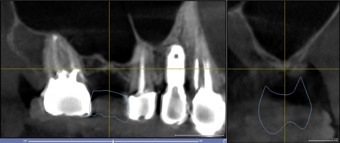
Figure 1: Preoperative CBCT scanning images of site #3
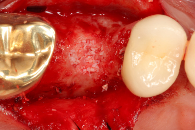
Figure 2: Occlusal view after completing the lifting procedure of the transcrestal sinus staging
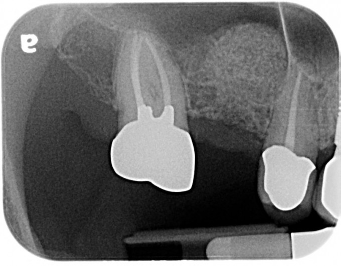
Figure 3: Periapical Radiography of the Transcrestal Breast Elevation Procedure
After a healing period without incident or 6 months, a radiographic evaluation of CT periapical and cone beam was compensated and implant planning was carried out in the SICAT 2.0 implant planning software (Figure 4). The digital implant plan was loaded to Sicat in Germany for the design of a digital guide, which was printed in 3D at home. The surgical steps of surgery of the second stage were the following:
1.) Preparation of a complete thickness of the Bucco-Linguistic wrap flap sites #2-4.
2.) Fully -guided placement or an Astra Ev S 4.2 x 9 mm implant (Figure 5).
3.) Minor additional oral increase with a mixture of a xenoinjut (Bio-Oss, Geistlich Pharma) and platelet-rich fibrin (PRF) survived with a long-lasting ossifying collagen membrane (Ossix Volumax, Dentspsplemax.
4.) Placement of a Transucose Healing Cover (Astra Ev Healdesign 5 x 4.5) and non -substantial suture around the healing cover with a non -resistant suture (5.0 Cytoplast, Osteogenics).
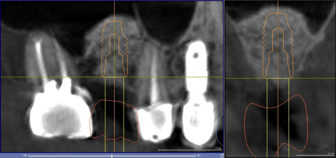
Figure 4: Second stage implant plan in sicat 2.0
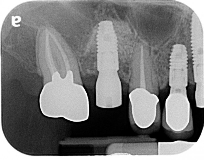
Figure 5: Immediate periapical radiography after implant
After a healing period without additional incidents of 3 months, an atlantis IO Flo Scankbody was inserted in the implant and a digital cache impression of Cerec of the implant was obtained and charged in Atlantis. Atlantis designed and manufactured a personalized titanium base and the corresponding E.Max crown was internally grinding from the central archive provided by Atlantis for an implant crown reticidated by final screw (Figs. 6-8).
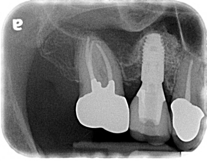
Figure 6: final periapical radiography or restored implant
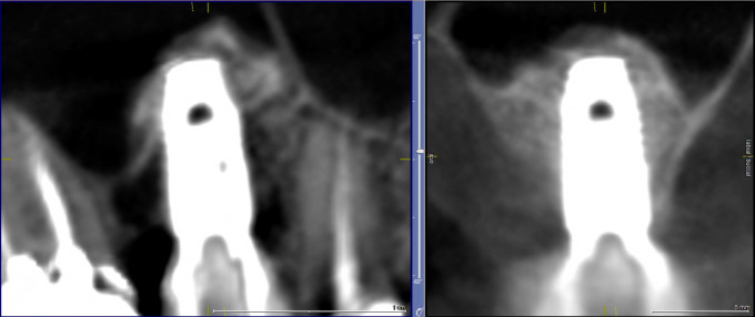
Figure 7: Final postoperative or implantation CBCT scanning
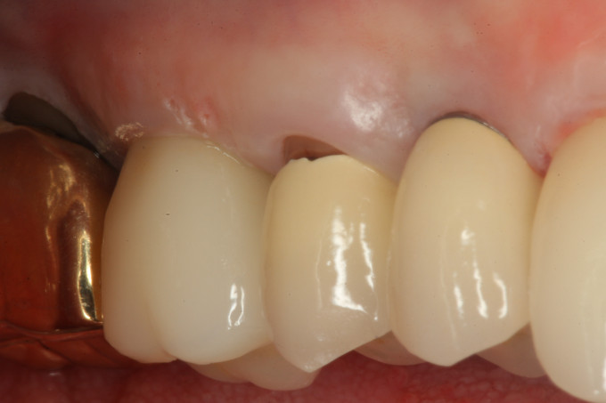
Figure 8: Final clinical view of the restored implant
The staging sinus elevator provides an attractive minimally invasive alternative to the traditional sine elevation technique. The key to the successful implementation of this technique is to maintain the integrity of the Schneiderian membrane to guarantee the containment of bone graft. The surgeon must be prepared to resort to a traditional sine elevation procedure in case a membrane drilling is produced to repair membrane drilling and increase the breast at the same time.