


Posted 1381 days ago
By Karyn M. Halpern DMD, MS
Patient presentation
TO Healthy male of 45 years presented a hygiene visit and an exam without complaints. After a clinical and radiographic examination, a great proximal caries adjacent to a occlusal-linguistic resin restoration was observed in the tooth #3. In addition, the tooth n. ° 2, diagnosed with occlusal-linguistic and fissure caries, as well as the proximal mesial caries (Figures 1 and 2, then).
YOUPut shown and informed of the findings, it was recommended to restore the great injury in the tooth #3 with a unique Cerec® filling and restore the smallest lesion in tooth #2 with a direct resin restoration.
![]()
Figure 1: Tooth #3 failing in occlusal-linguistic resin and a great distal recurring decomposition

![]()
Figure 2: Radiographic proximal caries in the tooth #2 mesial and #3 distal
Technique
The resin restoration was withdrawn in tooth #3 and the extensive distal decomposition was excavated. The distal lingual cusp restricted and thin was prepared for cusp coverage. Caries detector was applied to help verification or complete caries Elimination (Figure 3, below). The caries of well and fissure was removed in the tooth #2, as well as the decomposition in the mesial wall that was directly accessed. A G-AENIAL ™ Universal Flowable compound was used to restore #2 mesial, occlusal-lingüe, and also placed on the pulp floor of the preparation of the tooth #3.
As with all CEREC® restorations, the key to success is reduced to preparation. For partial coverage of coverage, the preparation must have drawn without companies, especially in the interproximal box. He Internal line angles must be round and smooth, with straight output walls in the interproximal.
![]()
Figure 3: All of preparation of the N #3 propellation and the application of the caries detector to help In caries elimination
After the compound Restoration in n. ° 2 and preparation in n. ° 3 were completed, the lower jaw, the upper jaw and the oral bite were recorded using the CERREC® priority in the acquisition phase. Virtual models and margins were created in virtual preparation were created using the automatic margin search engine using the CERREC® 5.2 software (Figure 4). Once the design was completed, the restoration of the line was manufactured using a Cerasmart® 270 A2 LT block with MCXL of Syrona Dentsply. He quickly and predictedly bothers without any marginal chipping (Figure 5). It also saves time since you don’t need to shoot.
![]()
Figure 4: Margination using the automatic margin search engine
![]()
Figure 5: Grew margins without marginal chief
Once milling, the swimsuit was deleted. The restoration sat then, verifying the margins closed and The contact was marked with tooth thread. The restoration was then characterized by using the red brown stain Optiglaze ™ in the occlusal fissures followed by a transparent layer of Optiglaze ™.
To deliver the Enter and for the final placement, the G-CEM One ™ self-adhesive resin cement was used in “adhesive cement mode” combining it with the adhesive improvement printing G-CEM One ™ adhesive (“aep”). AEP accelerates the chemical cure of cement to allow optimal link. It allows extremely easy cleaning, which makes it even more advantageous..
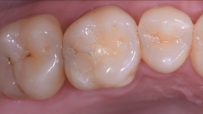
Once linked to the tooth, Restoration margins are mixed with the structure of the surrounding tooth (Figure 6, below).
![]()
Figure 6: Occlusal Vista of Final sentence CARASMART®270 A2 LT Restoration #3 after cementation
Advantages
This clinical The case demonstrates how a Partial coverage CEREC® undulating It can be designed, manufacturedand placed on a single visit using CARASMART® 270 and G-CEM One ™ Resin cement. CARASMART®270 factories wonderfully without shaking and saves a significant chair time, since it is not necessary to shoot. It can be easily characterized by Optiaze ™ cSmell to match the existing aesthetics or simply polished and placed. When combined with G-Cem one™ with AEPThe result is a strong and aesthetic restoration that combines harvester with the structure of the surrounding tooth.


